
The cochlea is an important part of the ear for hearing. It is sound in the inner ear and is believed to form during the embryonic period, as early as 4 weeks post gestation. It is fully formed by 18 weeks and this process is controlled by a gene known as SOX2. Mutations of this gene are strongly associated with hearing loss.
If you suffer from severe hearing loss, you may be offered a cochlear implant. This is similar to a hearing aid but can help much more significantly. If you are worried about your hearing you should visit an audiologist as soon as possible.
Structure and Anatomy
The cochlea is a small, spiral-shaped bone that is used to transduce a number of different sound frequencies. This is the process where the ear converts sound waves into electrical impulses. These are then sent to the brain which allows us to interpret them as sound.
The width of the cochlea is around 10 mm, and if it were to be unwound, the cochlea would measure around 35 mm long.
The spiral is filled with 2 different types of fluid, known as perilymph and endolymph. Perilymph is an extracellular fluid that is similar in makeup to cerebrospinal fluid and blood plasma. Endolymph differs as it has a high concentration of potassium ions and a low concentration of sodium ions. Endolymph is also known as Scarpa fluid and is clear.
The cochlea is split up into 3 distinct chambers. These are the scala vestibuli, scala media, and the scala tympani. The scala vestibuli, or vestibular duct, is the top area in the cochlear duct. This is filled with perilymph and is designed to conduct the sound vibrations into the cochlear duct.
The scala media, or cochlear duct, is found in the center of the other 2 chambers. It is full of endolymph. It is held in place with Reissner’s membrane, a very veiny stria vascularis, and the basilar membrane. Stria vascularis, or the spiral ligament, forms the outer wall of the cochlear duct. It has a lot of capillaries and tiny blood vessels and is designed to produce endolymph.
The scala tympani, or tympanic duct, is the lower area of the cochlea, underneath the basilar membrane. It is filled with perilymph and extends to the helicotrema where it becomes a vestibular duct. This chamber is designed to sense pressure changes caused by sound.
The cochlea also contains a number of minuscule hair cells specific to the organ of Corti. These are integral to hearing. These hair cells do not regenerate when lost and can easily become damaged by loud noises, trauma, or health conditions. This means that if you begin to lose these hairs you will suffer from permanent sensorineural hearing loss.
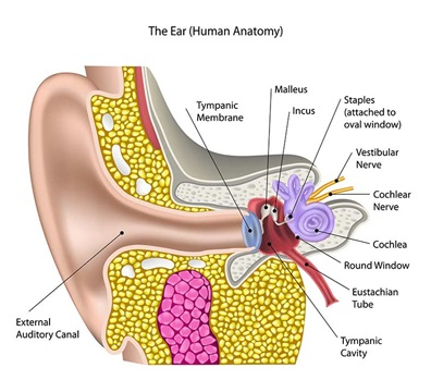
Where Is The Cochlea?
The cochlea is found in the inner ear. It is one of two main structures found here, the other being the semicircular canals, also known as the labyrinthine.
To find the cochlea, you first need to locate the inner ear. This is behind the eardrum, next to the inner ear. Once you have reached the eardrum, you will find a series of tiny bones known as the ossicles. These are integral to hearing.
Behind the ossicles is the oval window, and then the semicircular canals. These are filled with fluid, which is what helps you to retain your sense of balance. Adjacent to this you will find the round window, behind which is the cochlea.
What Is The Function of the Cochlea?
The cochlea is absolutely vital to hearing. It is used to transform the vibrations of the lymph fluids and their associated structures into an electrical signal that can be transmitted along nerves and into the brain.
This happens at the organ of Corti. This is a collection of sensory cells (the tiny hairs) that change the mechanical vibration to electrical impulses. These impulses are sent along the auditory nerve into the brain.
The vibrations force the hair cells to move about. As they move, the cells brush their stereocilia (the small hair-like projections on the upper side of the cells) along the tectorial membrane.
This movement depolarizes the attached nerve fibers. This is a change in the electrolyte balance of the fluid surrounding the cells, and what creates the electrical impulse.
Cochlear Implants
This is a small electronic device that can be implanted into the skull to enhance the hearing abilities of the wearer. It will electrically stimulate the cochlear nerve to allow the hearing to be restored. They are more invasive than traditional hearing aids and so will not be used unless the traditional versions prove ineffective.
There are 2 parts to a cochlear implant, internal and external elements. The external element sits behind the user’s ear and contains a microphone to pick up sounds from around you. This is then processed and transmitted via a wire to the internal element.
The internal element is located behind the ear too, but subcutaneously (underneath the skin). This is connected to the cochlea with a thin wire and a number of small electrodes. These send signals to the cochlear nerve, which in turn sends signals to the brain.
You will need to undergo therapy to ensure your cochlear implant works as efficiently as possible for you. Normal hearing will never be completely restored, but these implants can go a long way towards making your life easier. You will need to practice regularly to get the most out of them.
Cochlear implants are not the same as a hearing aid. Hearing aids simply amplify sounds, whereas cochlear implants can help to improve the sound quality, in turn improving your speech understanding.
Cochlear implants can be given to people of all ages. Early intervention is key as the hearing will continue to deteriorate over time. This means that sometimes cochlear implants are given to infants and toddlers to improve their hearing before they suffer too much of a loss. This can also help them with their speech development.